Monday, 11 December 2023

Understanding Wrist Pain on Top of Your Hand

What is a Wrist Pain on Top of Your Hand?

Wrist Pain on Top of Your Hand

Pain on the top of your wrist, just beneath the base of your fingers, can disrupt your daily life, making even simple tasks like gripping objects or typing feel uncomfortable. While causes vary, understanding the potential sources of this pain can guide you towards effective treatment and relief. This article delves into the common culprits behind wrist pain on top of the hand, exploring potential contributors such as:

Overuse injuries: Repetitive motions like typing, playing sports, or using tools can strain tendons and muscles, leading to inflammation and pain.

Carpal tunnel syndrome: This condition compresses the median nerve, causing tingling, numbness, and weakness in the thumb, index, and middle fingers. Pain can radiate up the wrist and be felt on the top.

Tendonitis: Inflammation of the tendons that connect muscles to bones can cause localized pain and tenderness on the top of your wrist.

Ganglion cysts: These fluid-filled sacs can form on top of the wrist, causing pain and limited movement.

Arthritis: Osteoarthritis and rheumatoid arthritis can damage the joints within the wrist, leading to pain, stiffness, and inflammation.

Fractures: A break in one of the wrist bones can cause pain on the top of the hand, accompanied by swelling and bruising.

This article will provide a comprehensive overview of each potential cause, including symptoms, diagnosis, and treatment options. By understanding the source of your wrist pain, you can take proactive steps toward recovery and prevent future discomfort.

Causes

Causes of Wrist Pain on Top of Your Hand:

Several factors can contribute to pain on the top of your wrist, ranging from overuse injuries to underlying conditions. Here's a closer look at some of the most common causes:

1. Overuse Injuries:

Repetitive Strain Injury (RSI): Repetitive motions involved in typing, playing sports, using tools, or gardening can strain tendons and muscles, leading to inflammation and pain on the top of your wrist.

Tenosynovitis: This condition inflames the lining of the tendon sheath, causing pain, friction, and difficulty moving the wrist.

De Quervain's Tenosynovitis: This specific type of tenosynovitis affects the tendons that control thumb movement, causing pain on the side of the wrist at the base of the thumb.

2. Carpal Tunnel Syndrome:

The median nerve travels through a narrow tunnel in the wrist. When this space compresses the nerve, it can cause tingling, numbness, and weakness in the thumb, index, and middle fingers. This pain can sometimes radiate up the wrist and be felt on the top.

3. Tendonitis:

Tendons are fibrous cords that connect muscles to bones. When these tendons become inflamed, they can cause pain, tenderness, and swelling at the affected site. Tendonitis affecting the wrist extensor tendons, located on the top of the wrist, can manifest as pain on the top of the hand.

4. Ganglion Cysts:

These fluid-filled sacs often form on the top of the wrist, causing pain, tenderness, and limited movement. While they may not always cause pain, pressure on the cyst or nearby nerves can trigger discomfort.

5. Arthritis:

Both osteoarthritis (wear-and-tear) and rheumatoid arthritis (an autoimmune disease) can affect the joints within the wrist, causing pain, stiffness, inflammation, and swelling. The pain can be localized on the top of the wrist and worsen with movement.

6. Fractures:

A break in one of the wrist bones, such as the scaphoid or lunate, can cause immediate pain on the top of the hand, accompanied by swelling, bruising, and difficulty moving the wrist.

7. Other Causes:

Although less common, other potential causes of wrist pain on top of the hand include:

Bursitis: Inflammation of the bursa, a fluid-filled sac that cushions bones and tendons.

Nerve entrapment: Compression of nerves passing through the wrist can cause pain, tingling, and numbness.

Infections: Bacterial or viral infections in the wrist joint can cause pain, swelling, and warmth.

Tumors: While rare, benign or malignant tumors can develop in the wrist, causing pain and other symptoms.

It's crucial to remember that this list is not exhaustive, and it's always best to consult a healthcare professional for diagnosis and proper treatment of wrist pain.

Anatomy of the Wrist Joint

The wrist joint, also known as the radiocarpal joint, is a complex articulation that allows for a wide range of motion in the hand. It is formed by the distal ends of the radius and ulna, two forearm bones, and the proximal row of carpal bones, eight small bones that form the wrist.

Here is a breakdown of the key anatomical structures of the wrist joint:

Bones:

Radius: The larger of the two forearm bones, the radius forms the main articulation with the carpal bones

Ulna: The smaller of the two forearm bones, the ulna provides stability to the wrist joint.

Carpal bones: There are eight carpal bones arranged in two rows. The proximal row includes the scaphoid, lunate, triquetral, and pisiform bones. The distal row includes the trapezium, trapezoid, capitate, and hamate bones.

Metacarpal bones: These five bones connect the wrist to the fingers.

Joints:

Radiocarpal joint: This is the main joint of the wrist, formed by the articulation of the radius with the scaphoid, lunate, and triquetral bones.

Midcarpal joint: This joint connects the two rows of carpal bones.

Carpometacarpal joints: These joints connect the carpal bones to the metacarpal bones.

Ligaments:

Collateral ligaments: These ligaments provide stability to the wrist joint on the radial and ulnar sides.

Dorsal and palmar ligaments: These ligaments support the wrist joint on the top and bottom, respectively.

Intercarpal ligaments: These ligaments connect the carpal bones to each other.

Cartilages:

Articular cartilage: This smooth cartilage covers the ends of the bones within the wrist joint and helps to reduce friction.

Triangular fibrocartilage complex (TFCC): This fibrocartilage pad helps to stabilize the ulnar side of the wrist joint.

Nerves and arteries:

Median nerve: This nerve passes through the carpal tunnel in the wrist and provides sensation to the thumb, index, middle, and half of the ring finger.

Ulnar nerve: This nerve travels along the ulnar side of the wrist and provides sensation to the little finger and half of the ring finger.

Radial artery: This artery supplies blood to the wrist and hand.

Understanding the anatomy of the wrist joint is essential for diagnosing and treating wrist pain, injuries, and conditions.

Sign and Symptoms

Pain on the top of your wrist can manifest in various ways, depending on the underlying cause. Here are some common symptoms and signs to watch for:

Pain:

Aching, throbbing, or sharp pain on the top of the hand, typically localized to a specific area.

Pain that worsens with certain activities, such as typing, gripping objects, or performing repetitive motions.

Difficulty moving the wrist due to pain and stiffness.

Swelling:

Noticeable swelling on the top of the wrist, particularly around the affected area.

Tenderness to the touch, making it difficult to press on the painful area.

Limited movement:

Difficulty bending and extending the wrist due to pain and stiffness.

Reduced grip strength and difficulty holding objects.

Other symptoms:

Clicking or popping sounds in the wrist when moving it.

Numbness or tingling in the thumb, index, middle, or ring finger (if the median nerve is compressed).

Weakness in the hand and fingers.

Difficulty writing or performing other fine motor tasks.

Here are some additional signs and symptoms that might indicate specific causes of wrist pain:

Carpal tunnel syndrome:

Numbness, tingling, or burning sensation in the thumb, index, middle, and half of the ring finger.

Difficulty gripping objects due to weakness in the hand.

Pain that radiates up the forearm.

Tendonitis:

Tenderness and swelling over the affected tendon.

Pain that worsens with specific movements that involve the affected tendon.

Ganglion cysts:

A visible, movable lump on the top of the wrist.

Pain that worsens when pressure is applied to the cyst.

Arthritis:

Stiffness and pain in the wrist, especially in the mornings or after periods of inactivity.

Difficulty making a fist or performing other gripping tasks.

Joint swelling and warmth.

Fractures:

Immediate pain and swelling after a fall or injury.

Difficulty moving the wrist and hand.

Deformity of the wrist.

It's important to note that this is not an exhaustive list, and individual symptoms may vary depending on the underlying cause of your wrist pain.

If you experience any of these symptoms, it's crucial to seek medical attention for diagnosis and proper treatment. Early diagnosis and treatment can help prevent complications and promote faster healing.

Differential Diagnosis

  • Repetitive Strain Injury (RSI)
  • De Quervain's Tenosynovitis
  • Carpal Tunnel Syndrome
  • Osteoarthritis Muscle Tendonitis
  • Ganglion Cysts
  • Wrist Osteoarthritis
  • Rheumatoid arthritis
  • Scaphoid fracture
  • Lunate fracture
  • Wrist Bursitis
  • Carpal Tunnel Syndrome

Diagnosis

Diagnosing Wrist Pain on Top of Your Hand

Diagnosing the exact cause of your wrist pain on top of your hand requires a comprehensive approach that involves a combination of medical history, physical examination, and diagnostic tests. Here's a breakdown of the diagnostic process:

1. Medical History:

Your doctor will inquire about your symptoms, including:

Location and intensity of the pain.

Onset and duration of the pain.

Activities that worsen the pain.

Any recent injuries or falls.

Any pre-existing conditions such as arthritis or diabetes.

Your doctor will also ask about your daily activities, occupation, and hobbies to identify potential causes related to repetitive motions or overuse.

2. Physical Examination:

Your doctor will examine your wrist, looking for:

Swelling.

Tenderness.

Redness.

Deformity.

Range of motion limitations.

They will also perform specific tests to assess for:

Phalen's test: This test involves flexing the wrists for a minute to see if it provokes numbness or tingling in the fingers, suggesting carpal tunnel syndrome.

Tinel's sign: This test involves tapping the median nerve at the wrist to see if it causes tingling or pain, also suggesting carpal tunnel syndrome.

Grip strength: Your doctor will measure your grip strength to identify any weakness that might indicate nerve compression or tendon problems.

3. Diagnostic Tests:

Depending on the suspected cause of your pain, your doctor may order additional tests, such as:

X-rays: These can reveal fractures, dislocations, or bone abnormalities.

MRI scans: These provide detailed images of soft tissues, such as tendons and ligaments, which can help diagnose tendonitis, ligament sprains, or ganglion cysts.

Nerve conduction studies: These tests measure the electrical activity of nerves to assess for nerve compression or damage.

Blood tests: These can help diagnose inflammatory conditions such as rheumatoid arthritis or gout.

Combining information from your medical history, physical examination, and diagnostic tests, your doctor will be able to pinpoint the cause of your wrist pain and recommend the most appropriate treatment plan.

Here are some additional points to remember:

Early diagnosis is crucial for effective treatment and preventing further complications.

The specific diagnostic tests ordered will vary depending on your individual case and symptoms.

It's important to communicate openly with your doctor about any questions or concerns you have regarding the diagnostic process.

By actively participating in your diagnosis and treatment, you can optimize your recovery and improve your wrist pain.

Treatment of Wrist Pain on Top of Your Hand

Depending on the underlying cause and severity of your wrist pain, your doctor will recommend the most effective treatment plan. Here's an overview of common treatment options for wrist pain on top of your hand:

1. Non-surgical Treatments:

Rest: Avoiding activities that aggravate your wrist is crucial for promoting healing. This may involve taking a break from work, hobbies, or sports that involve repetitive hand motions.

Ice: Applying ice packs wrapped in a towel to the affected area for 20-minute intervals, several times a day, can help reduce inflammation and pain.

Compression: Wearing a wrist splint or brace can help support the wrist and reduce pain and swelling.

Elevation: Keeping your wrist elevated above heart level can help reduce swelling and promote healing.

Pain relievers: Over-the-counter pain medications like ibuprofen or acetaminophen can help manage pain and inflammation.

Physical therapy: Exercises designed to strengthen and stretch the muscles and tendons around the wrist can help improve flexibility, range of motion, and prevent future injuries.

Occupational therapy: This can help you modify your work or daily activities to avoid aggravating your wrist pain.

Injections: Corticosteroid injections can be used to reduce inflammation and pain in cases of carpal tunnel syndrome or tendonitis.

2. Surgical Treatments:

In some cases, surgery may be necessary if non-surgical treatments fail to provide adequate relief. Examples of surgical procedures include:

Carpal tunnel release: This surgery involves cutting the ligament that compresses the median nerve in the wrist.

Tendon repair: This surgery may be needed to repair torn tendons caused by injuries or overuse.

Ganglion cyst removal: This surgery involves removing the ganglion cyst from the wrist.

The specific treatment recommended for your wrist pain will depend on the underlying cause, severity of symptoms, and your individual needs. It's important to discuss all treatment options with your doctor and ask any questions you may have.

Here are some additional points to remember:

It is crucial to follow your doctor's instructions carefully to maximize the effectiveness of your treatment plan.

The recovery time for wrist pain can vary depending on the severity of the condition and the chosen treatment.

Physical therapy plays a vital role in rehabilitation, helping you regain strength, flexibility, and function in your wrist.

Maintaining good posture and avoiding repetitive hand motions can help prevent future wrist pain.

If your wrist pain persists or worsens despite treatment, consult your doctor for further evaluation and possible adjustments to your treatment plan.

Physical Therapy for Wrist Pain on Top of Your Hand

Physical therapy plays a crucial role in treating and managing wrist pain on top of your hand. A physical therapist can assess your specific condition, develop a personalized exercise program, and guide you through proper technique to maximize your recovery and prevent future injuries. Here's an overview of the types of physical therapy treatments commonly used for wrist pain:

1. Manual Therapy:

Joint mobilization and manipulation: These techniques gently move the wrist joints to improve range of motion, reduce stiffness, and alleviate pain.

Soft tissue massage: This can help reduce muscle tension, improve blood circulation, and promote healing.

Trigger point therapy: This technique focuses on releasing tight knots in muscles that can contribute to pain and discomfort.

2. Strengthening Exercises:

Isometric exercises: These involve contracting muscles without joint movement, helping to build strength and endurance in the wrist muscles.

Isotonic exercises: These involve moving the wrist through its range of motion while holding weights or resistance bands, further strengthening the muscles.

Eccentric exercises: These involve lengthening the muscles under tension, which can help improve flexibility and prevent future injuries.

3. Stretching Exercises:

Stretching the muscles and tendons around the wrist can help improve range of motion, flexibility, and reduce pain. Common stretches include:

Wrist flexor stretch: Pull the fingers back towards the forearm to stretch the muscles on the palm side of the wrist.

Wrist extensor stretch: Bend the hand back towards the forearm to stretch the muscles on the top of the wrist.

Radial and ulnar deviation stretches: Tilt the hand sideways to stretch the muscles on the thumb and little finger sides of the wrist.

Other Modalities:

Ultrasound: This uses sound waves to generate heat deep within the tissues, which can help reduce pain and inflammation.

Electrical stimulation: This uses electrical currents to stimulate muscles and nerve pathways, which can help improve pain management and muscle strength.

Heat therapy: Applying heat packs to the affected area can help increase blood flow and reduce pain and stiffness.

Ice therapy: Applying ice packs to the affected area can help reduce inflammation and pain, especially after exercise or activity.

Your physical therapist will tailor your treatment program to your specific needs and condition. They will provide you with instructions on how to perform each exercise correctly and will monitor your progress throughout your treatment journey.

Here are some additional benefits of physical therapy for wrist pain:

  • Improves grip strength and dexterity
  • Reduces pain and inflammation
  • Enhances range of motion and flexibility
  • Helps prevent future injuries
  • Educates you on proper wrist mechanics and care
  • Provides pain management strategies

If you are experiencing wrist pain on top of your hand, consulting a physical therapist can be an effective way to manage your symptoms and regain full use of your hand.

How to Prevent Wrist Pain on Top of Your Hand?

Taking proactive steps to prevent wrist pain can significantly improve your hand health and avoid future discomfort. Here are some effective strategies:

1. Ergonomics:

Maintain proper posture: Sit upright with your shoulders relaxed and your elbows bent at 90 degrees when working at a desk.

Adjust your workstation: Ensure your chair height and keyboard position allow your wrists to remain straight while typing.

Use ergonomic tools: Consider using a wrist rest, keyboard with split keys, and a vertical mouse to promote comfortable hand positions.

2. Avoid Repetitive Motions:

Take frequent breaks: When engaging in repetitive hand motions, such as typing or using tools, take regular breaks to stretch and rest your hands.

Change positions: Vary your hand and wrist positions throughout the day to avoid overusing specific muscles and tendons.

Learn proper technique: If you engage in activities that involve repetitive hand motions, such as playing sports or playing musical instruments, ensure you use proper technique to minimize strain on your wrists.

3. Strengthen and Stretch:

Perform regular wrist exercises: Include exercises that target the muscles and tendons in your forearm and hand to build strength and flexibility.

Stretch your wrists: Regularly stretch the muscles and tendons in your hands and forearms to maintain good range of motion and prevent stiffness.

Warm up before activities: Before engaging in activities that involve repetitive hand motions, perform a gentle warm-up routine to increase blood flow and prepare your muscles.

4. Listen to Your Body:

Pay attention to pain: If you experience pain or discomfort in your wrist, stop the activity you're doing and rest your hand.

Apply ice: Ice packs can help reduce inflammation and pain, especially after activity.

Elevate your wrist: Keeping your wrist elevated above your heart can help reduce swelling and promote healing.

5. Lifestyle Modifications:

Maintain a healthy weight: Excess weight can put additional stress on your wrists. Maintaining a healthy weight can help reduce strain and prevent injuries.

Eat a balanced diet: Eating a diet rich in vitamins and minerals can help ensure your body has the necessary nutrients to maintain strong bones and healthy joints.

Stay hydrated: Drinking plenty of water helps keep your joints lubricated and reduces inflammation.

Avoid smoking: Smoking can damage your blood vessels and limit blood flow to your hands and wrists, increasing your risk of pain and injury.

By following these preventative measures, you can significantly reduce your risk of developing wrist pain on top of your hand and maintain optimal hand health.

Remember, if you experience persistent or severe wrist pain, consult a doctor for diagnosis and appropriate treatment.

Conclusion: Taking Charge of Your Wrist Health

Wrist pain on top of your hand can significantly impact your daily life, making even simple tasks challenging. However, understanding the potential causes, available treatments, and preventative measures empowers you to take charge of your hand health and achieve optimal pain management.

Here's a summary of the key takeaways:

Causes: Several factors can contribute to wrist pain, including overuse injuries, carpal tunnel syndrome, tendonitis, ganglion cysts, arthritis, and fractures.

Symptoms: Common symptoms include pain, swelling, stiffness, limited movement, tingling, numbness, and weakness.

Diagnosis: A comprehensive approach involving medical history, physical examination, and diagnostic tests is crucial for accurate diagnosis.

Treatment: Options range from non-surgical methods like rest, ice, pain relievers, physical therapy, and occupational therapy to surgical interventions in severe cases.

Prevention: Ergonomics, avoiding repetitive motions, strengthening and stretching exercises, listening to your body, and maintaining a healthy lifestyle can significantly reduce your risk of developing wrist pain.

By actively participating in your diagnosis, treatment, and prevention efforts, you can manage your wrist pain effectively and maintain optimal hand function for a pain-free and fulfilling life.

Monday, 27 March 2023

Multiple Sclerosis: Cause, Symptoms, Treatment, Exercise

What is a Multiple Sclerosis?

Multiple Sclerosis
Multiple Sclerosis

Multiple sclerosis (MS) is a chronic autoimmune disease that affects the central nervous system (CNS), which includes the brain, spinal cord, and optic nerves. MS is characterized by inflammation and damage to the myelin sheath that surrounds and protects nerve fibers in the CNS, leading to a variety of neurological symptoms and disability.

MS can affect people of any age, but it is most commonly diagnosed in young adults between the ages of 20 and 40. The exact cause of MS is not fully understood, but it is thought to involve a combination of genetic and environmental factors.

Symptoms of MS can vary widely depending on the location and extent of damage in the CNS, but common symptoms include fatigue, muscle weakness or spasms, numbness or tingling, vision problems, balance and coordination problems, and cognitive impairment.

There is currently no cure for MS, but there are a number of treatments available that can help manage symptoms, slow disease progression, and improve quality of life for people with MS. These treatments may include medications, physical therapy, occupational therapy, and other supportive therapies.

Related Anatomy

To understand multiple sclerosis (MS), it's helpful to have a basic understanding of the anatomy of the central nervous system (CNS), which includes the brain, spinal cord, and optic nerves.

The brain is the most complex organ in the body, and it is responsible for controlling and coordinating all of our bodily functions. It is composed of three main parts: the cerebrum, the cerebellum, and the brainstem.

The cerebrum is the largest part of the brain and is divided into two hemispheres. It is responsible for conscious thought, perception, and voluntary movement.

The cerebellum is located at the base of the brain and is responsible for coordinating movement and balance.

The brainstem connects the brain to the spinal cord and is responsible for controlling many of our automatic functions, such as breathing and heart rate.

The spinal cord is a long, thin structure that runs from the brainstem down through the center of the back. It is responsible for transmitting sensory information from the body to the brain and sending motor signals from the brain to the muscles and organs.

The optic nerves are a pair of nerves that transmit visual information from the eyes to the brain.

In MS, the immune system mistakenly attacks and damages the myelin sheath that surrounds and protects nerve fibers in the CNS, leading to a range of neurological symptoms and disability. This damage can occur in various parts of the CNS, resulting in a variety of symptoms depending on the location and extent of the damage.

What are the causes of Multiple Sclerosis?


The exact cause of multiple sclerosis (MS) is not fully understood, but it is believed to be a complex interplay between genetic and environmental factors.

  • Genetic factors: Although MS is not directly inherited, there is a genetic component to the disease. Studies have shown that certain genetic variations are associated with an increased risk of developing MS. However, having these genetic variations does not necessarily mean a person will develop the disease.
  • Environmental factors: There are a number of environmental factors that have been linked to an increased risk of developing MS. These include:
  • Vitamin D deficiency: There is evidence to suggest that low levels of vitamin D may increase the risk of developing MS.
  • Smoking: Smoking has been shown to increase the risk of developing MS, as well as the severity of the disease.
  • Infections: Certain infections, such as the Epstein-Barr virus, have been linked to an increased risk of developing MS.
  • Geographic location: MS is more common in certain geographic regions, particularly those further from the equator.
  • Autoimmune dysfunction: MS is considered to be an autoimmune disease, meaning that the immune system mistakenly attacks and damages the myelin sheath in the central nervous system.


Overall, it is likely that MS is caused by a combination of genetic and environmental factors that interact to trigger the disease in susceptible individuals. However, the precise mechanisms underlying the development of MS are still not fully understood, and ongoing research is focused on unraveling the complex factors involved.

Symptoms of Multiple Sclerosis

Multiple sclerosis (MS) can cause a wide variety of symptoms, which can vary greatly between individuals depending on the location and extent of damage in the central nervous system. Common symptoms of MS include:

  • Fatigue: This is one of the most common symptoms of MS and can be debilitating, affecting a person's ability to carry out daily activities.
  • Numbness or tingling: MS can cause sensations of numbness, tingling, or pins and needles in different parts of the body.
  • Vision problems: MS can cause a range of vision problems, including blurred vision, double vision, or even temporary loss of vision.
  • Muscle weakness or spasms: MS can cause muscle weakness or spasms, which can make it difficult to move or carry out everyday activities.
  • Balance and coordination problems: MS can affect a person's balance and coordination, making it difficult to walk or perform other coordinated movements.
  • Cognitive impairment: MS can cause problems with memory, attention, and other cognitive functions.
  • Bladder and bowel problems: MS can affect the function of the bladder and bowel, leading to urinary or fecal incontinence or difficulty emptying the bladder or bowel.
  • Sexual dysfunction: MS can cause sexual dysfunction in both men and women.

It's important to note that these symptoms can also be caused by other conditions, so it's important to consult with a healthcare professional if you experience any of these symptoms. Additionally, symptoms of MS can vary in severity and frequency over time, and may come and go or worsen during periods of stress or illness.

Risk factor

There are a number of factors that can increase a person's risk of developing multiple sclerosis (MS), including:

  • Age: MS can occur at any age, but it most commonly affects people between the ages of 20 and 40.
  • Gender: MS is more common in women than men.
  • Family history: Having a close relative (such as a parent or sibling) with MS increases a person's risk of developing the disease.
  • Ethnicity: MS is more common in people of European descent than in other ethnic groups.
  • Geographic location: MS is more common in certain geographic regions, particularly those further from the equator.
  • Vitamin D deficiency: There is evidence to suggest that low levels of vitamin D may increase the risk of developing MS.
  • Smoking: Smoking has been shown to increase the risk of developing MS, as well as the severity of the disease.
  • Infections: Certain infections, such as the Epstein-Barr virus, have been linked to an increased risk of developing MS.


It's important to note that having one or more of these risk factors does not necessarily mean a person will develop MS, and many people with MS do not have any known risk factors. However, being aware of these risk factors can help people understand their individual risk and take steps to reduce their risk where possible (such as maintaining a healthy lifestyle and avoiding smoking).

Complications

Multiple sclerosis (MS) can lead to a number of complications, some of which can be quite serious. The specific complications that can occur vary depending on the location and extent of damage in the central nervous system, as well as other factors such as the severity of the disease and the individual's overall health.

Here are some common complications associated with MS:

  • Disability: Over time, MS can cause significant disability, such as difficulty with walking or other daily activities.
  • Depression and anxiety: MS can increase the risk of depression and anxiety, which can worsen overall quality of life.
  • Cognitive impairment: MS can cause problems with memory, attention, and other cognitive functions.
  • Bowel and bladder problems: MS can affect the function of the bladder and bowel, leading to urinary or fecal incontinence or difficulty emptying the bladder or bowel.
  • Respiratory problems: In some cases, MS can lead to respiratory problems, such as difficulty breathing or pneumonia.
  • Seizures: MS can increase the risk of seizures in some individuals.
  • Pain: MS can cause pain, including headaches, muscle pain, and nerve pain.
  • Sexual dysfunction: MS can cause sexual dysfunction in both men and women.

Increased risk of infections: People with MS may be at increased risk of infections, particularly if they are taking immunosuppressive medications to manage the disease.

It's important for individuals with MS to work closely with their healthcare team to manage symptoms and prevent complications. In some cases, medications, rehabilitation, and lifestyle changes may be recommended to help manage MS and prevent complications.

Differential Diagnosis

There are several conditions that may have symptoms similar to multiple sclerosis (MS) and can be considered in a differential diagnosis. Here are some examples:

  • Neuromyelitis optica (NMO): NMO is a rare autoimmune disorder that affects the optic nerves and spinal cord, and can cause symptoms similar to MS. However, NMO tends to cause more severe and rapid vision loss than MS, and may affect the spinal cord in a different pattern than MS.
  • Acute disseminated encephalomyelitis (ADEM): ADEM is a rare inflammatory condition that can cause symptoms similar to MS, including vision problems, weakness, and numbness. However, ADEM tends to develop more rapidly and typically follows an infection or vaccination.
  • Lyme disease: Lyme disease is a bacterial infection that can cause neurological symptoms similar to MS, including weakness, numbness, and difficulty with coordination. Lyme disease can be diagnosed with a blood test and treated with antibiotics.
  • Sarcoidosis: Sarcoidosis is a rare inflammatory condition that can affect multiple organs, including the nervous system. Neurosarcoidosis can cause symptoms similar to MS, including weakness, numbness, and difficulty with coordination.
  • Vitamin B12 deficiency: Vitamin B12 deficiency can cause neurological symptoms similar to MS, including weakness, numbness, and difficulty with coordination. A blood test can diagnose a vitamin B12 deficiency, and the condition can be treated with vitamin B12 supplements.

It's important to note that these conditions are rare and MS is the most common cause of symptoms such as those described above. However, a thorough medical evaluation and testing may be necessary to accurately diagnose MS or other conditions.

Diagnosis


The diagnosis of multiple sclerosis (MS) involves a combination of medical history, physical examination, and diagnostic testing. Here are the steps typically involved in the diagnostic process:

  • Medical history: The doctor will ask about the patient's symptoms and medical history, including any family history of MS or other autoimmune disorders.
  • Physical examination: The doctor will perform a neurological examination to assess the patient's strength, coordination, reflexes, and other neurological functions.
  • MRI: Magnetic resonance imaging (MRI) of the brain and spinal cord is often the primary diagnostic test for MS. The MRI can reveal areas of inflammation or damage (lesions) that are characteristic of MS.
  • Lumbar puncture: A lumbar puncture (also known as a spinal tap) may be performed to analyze the fluid that surrounds the brain and spinal cord for evidence of inflammation or abnormal immune system activity.
  • Evoked potentials: Evoked potentials are tests that measure the electrical activity of the brain in response to stimulation of the eyes, ears, or other sensory organs. These tests can help identify abnormalities in the nervous system that may be associated with MS.
  • Blood tests: Blood tests may be performed to rule out other conditions that can cause similar symptoms, such as Lyme disease or vitamin deficiencies.

The diagnosis of MS can be challenging, as symptoms can vary widely and other conditions may have similar symptoms. In some cases, additional testing or monitoring may be necessary to confirm a diagnosis. A neurologist or other healthcare provider with experience in diagnosing and treating MS can help guide the diagnostic process.

Treatment of Multiple Sclerosis

The treatment of multiple sclerosis (MS) aims to manage symptoms, slow disease progression, and improve quality of life. There is currently no cure for MS, but several medications and therapies are available to help manage the disease. The specific treatment plan for MS depends on the individual's symptoms, disease course, and other factors. Here are some examples of MS treatments:

  • Disease-modifying therapies (DMTs): DMTs are medications that can slow the progression of MS by reducing inflammation and suppressing the immune system. There are several types of DMTs available, including injectable, oral, and infusion therapies.
  • Symptom management: MS symptoms can be managed with a variety of medications and therapies, including corticosteroids for acute attacks, muscle relaxants for spasticity, and medications for fatigue, pain, and bladder and bowel problems.
  • Rehabilitation: Physical therapy, occupational therapy, and speech therapy can help individuals with MS improve their strength, mobility, and ability to perform daily activities.
  • Lifestyle changes: Lifestyle changes, such as regular exercise, a healthy diet, stress management, and smoking cessation, can help manage symptoms and improve overall health.
  • Complementary and alternative therapies: Some individuals with MS may benefit from complementary and alternative therapies, such as acupuncture, massage, and meditation. It's important to discuss any complementary or alternative therapies with a healthcare provider to ensure they are safe and effective.
  • Supportive care: MS can have a significant impact on an individual's quality of life, and supportive care can help address emotional, social, and practical challenges associated with the disease. Supportive care may include counseling, support groups, and vocational rehabilitation.

The treatment of MS is individualized and may involve a combination of medications, therapies, and lifestyle changes. It's important for individuals with MS to work closely with their healthcare team to develop a treatment plan that meets their specific needs and goals.

Physiotherapy treatment

Physical therapy treatment
, or physiotherapy, is a common treatment for multiple sclerosis (MS) that can help improve strength, flexibility, balance, and mobility. A physical therapist can work with individuals with MS to develop a customized exercise and rehabilitation program based on their specific needs and symptoms. Here are some examples of physiotherapy treatments for MS:

  • Stretching and range-of-motion exercises: Stretching and range-of-motion exercises can help improve flexibility and prevent muscle stiffness and contractures.
  • Strength training: Strength training exercises can help improve muscle strength and endurance, which can be especially beneficial for individuals with MS who experience muscle weakness or fatigue.
  • Balance and coordination training: Balance and coordination training can help reduce the risk of falls and improve overall mobility. This may include exercises such as standing on one leg or walking on uneven surfaces.
  • Gait training: Gait training involves exercises and techniques to improve walking ability, such as using assistive devices like canes or walkers, or practicing specific movements or techniques to improve balance and stability.
  • Aquatic therapy: Aquatic therapy involves exercises and rehabilitation activities performed in a pool or other aquatic environment. The buoyancy of the water can help reduce the impact of gravity on the body, making it easier to move and exercise.
  • Assistive devices: A physical therapist can help individuals with MS identify and obtain appropriate assistive devices, such as braces, orthotics, or mobility aids, to help improve mobility and reduce the risk of falls.

Physical therapy is an important component of MS treatment and can help individuals with MS maintain their independence and quality of life. It's important for individuals with MS to work closely with their healthcare team, including a physical therapist, to develop a comprehensive treatment plan that meets their specific needs and goals.

Exercise for Multiple Sclerosis


Regular exercise is an important part of managing multiple sclerosis (MS). Exercise can help improve strength, balance, flexibility, and mobility, as well as reduce fatigue and improve overall quality of life. However, it's important to work with a healthcare provider and/or a physical therapist to develop an exercise program that is safe and effective for individuals with MS. Here are some examples of exercises that may be beneficial for individuals with MS:

  • Aerobic exercise: Aerobic exercise, such as walking, cycling, or swimming, can help improve cardiovascular health and overall fitness. It's important to start slowly and gradually increase intensity and duration over time.
  • Strength training: Strength training exercises, such as weight lifting or resistance band exercises, can help improve muscle strength and endurance. It's important to use proper form and start with lighter weights or resistance bands.
  • Yoga: Yoga can help improve flexibility, balance, and relaxation. There are several modified yoga poses that are specifically designed for individuals with MS.
  • Tai Chi: Tai Chi is a gentle form of exercise that combines movement and meditation. It can help improve balance, flexibility, and relaxation.
  • Aquatic exercise: Aquatic exercise, such as swimming or water aerobics, can be a good option for individuals with MS who experience fatigue or difficulty with weight-bearing exercise.
  • Pilates: Pilates is a form of exercise that focuses on core strength and stability. It can help improve posture, balance, and coordination.

It's important to talk to a healthcare provider before starting an exercise program, especially if you have any medical conditions or concerns. A physical therapist can also help develop a customized exercise program that is safe and effective for individuals with MS.

How to Prevent Multiple Sclerosis?


At this time, there is no known way to prevent multiple sclerosis (MS), as the exact cause of the disease is not fully understood. However, there are some lifestyle factors that may help reduce the risk of developing MS or delay the onset of symptoms. Here are some steps that may help:

  • Maintain a healthy diet: Eating a healthy, balanced diet rich in fruits, vegetables, whole grains, and lean protein sources may help reduce inflammation and improve overall health.
  • Get enough vitamin D: Some studies suggest that low levels of vitamin D may be associated with an increased risk of MS. Getting enough vitamin D through sunlight exposure or supplements may help reduce the risk of MS or delay its onset.
  • Exercise regularly: Regular exercise can help improve overall health and may help reduce the risk of MS or delay its onset. Exercise can also help manage symptoms and improve quality of life for individuals with MS.
  • Manage stress: Stress may trigger or worsen MS symptoms in some individuals. Practicing stress-management techniques, such as deep breathing, meditation, or yoga, may help reduce stress and improve overall health.
  • Quit smoking: Smoking has been linked to an increased risk of MS and may worsen symptoms for individuals with the disease. Quitting smoking can help reduce the risk of MS or improve symptoms for those who already have the disease.

While there is no guaranteed way to prevent MS, adopting a healthy lifestyle and managing risk factors may help reduce the risk of developing the disease or delay its onset. It's also important to talk to a healthcare provider about any concerns or questions regarding MS and its prevention.

Conclusion


In conclusion, multiple sclerosis (MS) is a chronic and often disabling disease that affects the central nervous system. While the exact cause of MS is not fully understood, it is thought to be an autoimmune disorder in which the body's immune system attacks the myelin sheath that surrounds nerve fibers, causing damage and interference with nerve signals. The symptoms of MS can vary widely and may include fatigue, weakness, numbness, vision problems, balance and coordination issues, and cognitive impairment. 

There is no known cure for MS, but a variety of treatments are available to manage symptoms and slow the progression of the disease. In addition to medical treatments, lifestyle changes such as regular exercise, stress management, and a healthy diet may help improve symptoms and overall quality of life for individuals with MS. It's important to work with a healthcare provider and/or a physical therapist to develop a personalized treatment plan that is safe and effective for managing MS.

Wednesday, 22 March 2023

Lumbar Spondylolisthesis Cause, Symptoms, Treatment, Exercise

What is a Lumbar Spondylolisthesis?


Lumbar spondylolisthesis

Lumbar spondylolisthesis is a condition in which one vertebra in the lumbar spine (lower back) slides forward over the vertebra below it. This displacement can cause compression of the spinal nerves and lead to symptoms such as lower back pain, sciatica, numbness, tingling, and weakness in the legs.

The severity of lumbar spondylolisthesis is classified based on the degree of displacement of the affected vertebra. Grade 1 is mild and involves less than 25% slippage, while grade 2 involves 25% to 50% slippage, grade 3 involves 50% to 75% slippage, and grade 4 involves more than 75% slippage. In severe cases, the slippage can be so significant that the affected vertebra may slip completely off the vertebra below it.

Lumbar spondylolisthesis can be caused by various factors, including congenital abnormalities, degenerative changes in the spine, trauma, or repetitive stress injuries. Treatment options for lumbar spondylolisthesis depend on the severity of the condition and the presence of symptoms. Conservative treatment options may include rest, physical therapy, and pain management. In more severe cases, surgery may be required to stabilize the affected area and relieve pressure on the spinal nerves.

Related Anatomy

To understand lumbar spondylolisthesis, it is helpful to have a basic understanding of the anatomy of the lumbar spine.

The lumbar spine consists of five vertebral bones, labeled L1 to L5, which are located below the ribcage and above the sacrum. Each vertebra is separated by an intervertebral disc, which acts as a cushion and shock absorber. The intervertebral discs are made up of a tough outer layer called the annulus fibrosus and a gel-like inner core called the nucleus pulposus.

The vertebrae are held together by ligaments and muscles that provide stability to the spine. The ligaments include the anterior longitudinal ligament (located on the front of the vertebral bodies) and the posterior longitudinal ligament (located on the back of the vertebral bodies). The muscles of the lumbar spine include the erector spinae, quadratus lumborum, and psoas major, among others.

The spinal cord and spinal nerves run through the spinal canal, which is formed by the vertebrae. The spinal cord is a long, thin, tubular bundle of nerves that extends from the brainstem to the lower back. The spinal nerves branch out from the spinal cord and exit through small openings between the vertebrae, known as the neural foramina. These nerves transmit signals between the brain and the rest of the body.

Lumbar spondylolisthesis can affect any of the lumbar vertebrae and can cause compression or irritation of the spinal nerves that exit at that level. This can lead to symptoms such as pain, numbness, and weakness in the lower back and legs.

Causes of Lumbar Spondylolisthesis

Lumbar spondylolisthesis can have various causes, including:

  • Congenital: Some people are born with abnormalities in the structure of the vertebrae that can lead to spondylolisthesis.
  • Degenerative: As we age, the intervertebral discs lose moisture and become less flexible, leading to degenerative changes in the spine. These changes can cause the vertebrae to shift out of position.
  • Traumatic: A sudden impact or injury to the spine can cause the vertebrae to shift out of position.
  • Repetitive stress: Repetitive stress on the spine from activities such as weightlifting or gymnastics can cause spondylolisthesis, particularly in individuals who already have a congenital weakness in their vertebrae.
  • Isthmic: This type of spondylolisthesis occurs when there is a defect in the pars interarticularis, a small bony structure that connects the upper and lower parts of the vertebra. Isthmic spondylolisthesis is often caused by repetitive stress, such as from sports or certain occupations.
  • Pathologic: In some cases, spondylolisthesis may be caused by a pathologic condition, such as a tumor or infection, that weakens the vertebrae.
  • Postsurgical: Spondylolisthesis can occur as a complication of spine surgery.


The specific cause of lumbar spondylolisthesis can help guide treatment options and determine the prognosis for the condition. Treatment options may include rest, physical therapy, pain management, and in some cases, surgery.

Symptoms of Lumbar Spondylolisthesis

The symptoms of lumbar spondylolisthesis can vary depending on the degree of slippage and the extent of nerve compression. Some people with spondylolisthesis may have no symptoms at all, while others may experience the following:

  • Lower back pain: This is the most common symptom of lumbar spondylolisthesis. The pain may be dull or sharp and can be exacerbated by standing or walking for long periods of time.
  • Sciatica: This is a type of pain that radiates down one or both legs and is caused by irritation or compression of the sciatic nerve. Sciatica can cause pain, numbness, tingling, or weakness in the legs.
  • Stiffness: People with spondylolisthesis may feel stiffness in the lower back, which can make it difficult to move.
  • Weakness: Spondylolisthesis can cause weakness in the legs, which can affect balance and coordination.
  • Numbness or tingling: Compression of the spinal nerves can cause numbness or tingling in the lower back, legs, or feet.
  • Loss of bladder or bowel control: This is a rare symptom of lumbar spondylolisthesis, but it can occur in severe cases where the nerve compression is causing significant damage.


If you are experiencing any of these symptoms, it is important to see a doctor for an evaluation. A doctor can perform diagnostic tests and recommend appropriate treatment options based on the severity of your condition.

Risk factor

There are several risk factors that may increase a person's likelihood of developing lumbar spondylolisthesis. These include:

  • Age: Spondylolisthesis is more common in older adults, as the spine undergoes degenerative changes over time.
  • Gender: Spondylolisthesis is more common in women than in men.
  • Genetics: Some people may be born with a congenital weakness in the vertebrae that predisposes them to spondylolisthesis.
  • Occupation: Jobs that involve repetitive bending, twisting, or heavy lifting can increase the risk of spondylolisthesis.
  • Sports participation: Certain sports, such as gymnastics, weightlifting, and football, can put stress on the spine and increase the risk of spondylolisthesis.
  • Obesity: Excess body weight can put stress on the spine and increase the risk of spondylolisthesis.
  • Prior spinal surgery: Spondylolisthesis can occur as a complication of spinal surgery.
  • Spinal injury: A history of spinal injury or trauma can increase the risk of developing spondylolisthesis.


It is important to note that having one or more of these risk factors does not necessarily mean that a person will develop spondylolisthesis. However, it may be helpful to take steps to reduce the risk of developing spondylolisthesis, such as maintaining a healthy weight, practicing proper lifting techniques, and avoiding activities that put excessive stress on the spine.

What is the retro-listhesis of the Lumbar spine?

Retrolisthesis is a condition in which a vertebra in the spine shifts backward in relation to the vertebra below it. In the lumbar spine, retrolisthesis refers to a backward displacement of one vertebra in relation to the one below it. This can cause compression of the spinal nerves and lead to symptoms such as back pain, stiffness, and numbness or weakness in the legs.

Retrolisthesis can be caused by various factors, including degenerative changes in the spine, traumatic injury, or genetic conditions that affect the shape of the vertebrae. It is often seen in conjunction with other spinal conditions, such as spondylolisthesis or spinal stenosis.

Diagnosis of retrolisthesis typically involves imaging studies such as X-rays or MRI scans. Treatment depends on the severity of the condition and the symptoms it causes. Mild cases may be managed with conservative measures such as rest, physical therapy, and pain medications. In more severe cases, surgery may be necessary to correct the displacement and relieve pressure on the spinal nerves.

Differential Diagnosis

Lumbar spondylolisthesis can be diagnosed by a medical professional using imaging tests such as X-rays, MRI scans, or CT scans. However, it is important to consider other possible conditions that may have similar symptoms. Some possible differential diagnoses for lumbar spondylolisthesis include:

  • Lumbar spinal stenosis: This condition occurs when the spinal canal narrows and puts pressure on the spinal cord or nerve roots, causing similar symptoms to spondylolisthesis.
  • Herniated disc: A herniated disc occurs when the soft center of a spinal disc protrudes through a tear in the outer portion, which can cause compression of the spinal nerves and similar symptoms to spondylolisthesis.
  • Degenerative disc disease: This condition occurs when the intervertebral discs lose moisture and become less flexible, which can cause back pain and stiffness.
  • Osteoarthritis: This is a type of arthritis that can affect the spine and cause similar symptoms to spondylolisthesis.
  • Ankylosing spondylitis: This is a type of inflammatory arthritis that primarily affects the spine and can cause stiffness and pain.
  • Fibromyalgia: This is a chronic pain disorder that can cause widespread pain and fatigue, which can be mistaken for the symptoms of spondylolisthesis.


It is important to consult with a medical professional to obtain an accurate diagnosis and appropriate treatment plan for any spinal condition.

Diagnosis

The diagnosis of lumbar spondylolisthesis typically involves a medical history, physical examination, and imaging tests. Here are some common steps in the diagnostic process:

  • Medical history: The doctor will ask about your symptoms, medical history, and any risk factors for spondylolisthesis.
  • Physical examination: The doctor will perform a physical examination, which may include tests to assess your range of motion, muscle strength, and reflexes.
  • Imaging tests: Imaging tests such as X-rays, MRI scans, or CT scans may be used to confirm the diagnosis and determine the severity of the condition.
  • Electromyography (EMG): This test may be used to assess nerve function and determine whether nerve compression is contributing to your symptoms.
  • Bone scan: A bone scan may be ordered to determine whether there are any areas of increased bone activity, which can indicate stress fractures or bone abnormalities.

Once a diagnosis of lumbar spondylolisthesis is made, the doctor can determine the appropriate treatment plan based on the severity of the condition and the individual needs of the patient. Treatment options may include conservative measures such as physical therapy, pain medication, and activity modification, or in severe cases, surgical intervention may be necessary.

Treatment of Lumbar Spondylolisthesis


The treatment of lumbar spondylolisthesis depends on the severity of the condition and the individual needs of the patient. The treatment options can be broadly divided into two categories: conservative measures and surgical intervention.

Conservative Measures:


Physical therapy: Physical therapy can help strengthen the muscles in the back and abdomen to support the spine, and improve flexibility and range of motion.

  • Pain medication: Over-the-counter pain relievers, such as acetaminophen or nonsteroidal anti-inflammatory drugs (NSAIDs), can help alleviate pain and inflammation.
  • Activity modification: Avoiding activities that exacerbate symptoms, such as heavy lifting or high-impact sports, can help reduce pain and prevent further damage to the spine.
  • Bracing: Wearing a brace or support device can help stabilize the spine and provide pain relief.
  • Epidural steroid injections: Corticosteroids can be injected into the epidural space around the spinal cord to reduce inflammation and alleviate pain.

Surgical Intervention:

Laminectomy: This surgical procedure involves removing a portion of the bony arch (lamina) of the affected vertebra to relieve pressure on the spinal cord or nerve roots.

  • Fusion: In cases where the spondylolisthesis is severe or causing instability in the spine, a fusion procedure may be performed to stabilize the affected vertebrae and prevent further slippage.
  • Artificial disc replacement: This procedure involves replacing a damaged or degenerated disc with an artificial disc to maintain mobility in the affected area.
  • Vertebroplasty or kyphoplasty: These procedures involve injecting a cement-like material into a collapsed vertebra to stabilize it and alleviate pain.


It is important to consult with a medical professional to determine the most appropriate treatment plan for lumbar spondylolisthesis based on individual needs and circumstances.

Physiotherapy treatment for Lumbar Spondylolisthesis

Physical therapy can be an effective conservative treatment option for lumbar spondylolisthesis. A physiotherapist can design an individualized treatment plan to improve flexibility, strengthen muscles, reduce pain, and improve overall function. Here are some of the common physiotherapy treatments for lumbar spondylolisthesis:

  • Core strengthening exercises: These exercises focus on strengthening the muscles in the abdomen, lower back, and hips to support the spine and reduce strain on the affected area.
  • Stretching exercises: These exercises can improve flexibility in the muscles and joints of the back and hips, which can reduce pain and improve range of motion.
  • Manual therapy: Hands-on techniques such as massage, joint mobilization, and manual traction can help reduce pain and improve spinal alignment.
  • Posture education: Learning proper posture and body mechanics can help reduce strain on the spine and prevent further damage.
  • Electrical stimulation: This treatment involves the use of low-level electrical currents to stimulate the muscles and reduce pain.
  • Heat or ice therapy: Applying heat or ice to the affected area can help reduce inflammation and alleviate pain.
  • Education and advice: A physiotherapist can provide advice on modifications to activities and lifestyle changes that can help manage symptoms and prevent further damage to the spine.

It is important to consult with a physiotherapist to determine the most appropriate treatment plan for lumbar spondylolisthesis based on individual needs and circumstances.

Exercise for Lumbar Spondylolisthesis

Exercise can be an important component of a comprehensive treatment plan for lumbar spondylolisthesis. However, it is important to consult with a healthcare professional or a qualified physical therapist before starting any exercise program, as some exercises may exacerbate the condition or cause further damage. Here are some exercises that may be helpful for people with lumbar spondylolisthesis:

  • Pelvic tilt: Lie on your back with your knees bent and feet flat on the ground. Slowly tilt your pelvis forward and back, pressing your lower back into the ground and then arching it slightly. Repeat 10-15 times.
  • Cat-cow stretch: Start on your hands and knees, with your hands under your shoulders and your knees under your hips. Arch your back, tucking your chin to your chest, and then lower your back and lift your head, looking up towards the ceiling. Repeat 10-15 times.
  • Bird dog: Start on your hands and knees, with your hands under your shoulders and your knees under your hips. Reach your right arm forward and your left leg back, keeping your hips level. Hold for a few seconds, then switch sides. Repeat 10-15 times.
  • Wall squats: Stand with your back against a wall and your feet shoulder-width apart. Slowly slide down the wall into a squat, keeping your knees behind your toes. Hold for a few seconds, then slowly slide back up. Repeat 10-15 times.
  • Hamstring stretch: Lie on your back with your legs extended. Bend your right knee and place your foot flat on the ground. Slowly lift your left leg, keeping it straight, and grasp the back of your thigh with both hands. Hold for 30 seconds, then switch sides. Repeat 3-5 times on each side.


It is important to perform exercises under the guidance of a qualified professional to ensure proper form and technique, and to avoid exacerbating the condition or causing further damage to the spine.

Ergonomics


Ergonomics refers to the science of designing and arranging objects, equipment, and spaces in a way that maximizes efficiency, comfort, and safety. In the context of lumbar spondylolisthesis, ergonomics can be an important factor in reducing pain and preventing further damage to the spine.

Here are some ergonomic considerations to keep in mind:

  • Posture: Maintaining good posture can help reduce strain on the lower back. When sitting, make sure your feet are flat on the ground, your back is straight, and your shoulders are relaxed. When standing, distribute your weight evenly between your feet and avoid slouching.
  • Chair: Choose a chair that provides good lumbar support and is adjustable to your height. Use a cushion or rolled-up towel to provide extra support if needed.
  • Desk: Adjust the height of your desk so that your computer screen is at eye level, and your arms and wrists are in a comfortable position. Use a document holder to avoid straining your neck and back when reading or typing.
  • Keyboard and mouse: Use an ergonomic keyboard and mouse that provide wrist support and are positioned close to your body. Avoid resting your wrists on hard surfaces.
  • Lifting: Use proper lifting technique to avoid straining your back. Keep your back straight, bend your knees, and lift with your legs rather than your back. Avoid lifting objects that are too heavy or awkwardly shaped.

By implementing ergonomic principles, you can help reduce the strain on your lower back and prevent further damage to the spine. Consulting with an occupational therapist or ergonomic specialist can be helpful in designing a personalized ergonomic plan that meets your specific needs.

How to Prevent Lumbar Spondylolisthesis?

While it may not be possible to completely prevent lumbar spondylolisthesis, there are steps you can take to reduce your risk of developing this condition or prevent it from getting worse if you already have it. Here are some tips:

  • Exercise regularly: Strengthening the muscles that support your spine can help reduce the risk of spondylolisthesis. Consult with a healthcare professional or a qualified physical therapist to develop an exercise program that is safe and effective for your needs.
  • Practice good posture: Maintaining good posture can help reduce strain on your lower back. When sitting or standing, keep your back straight, shoulders relaxed, and feet flat on the ground.
  • Avoid overloading your spine: Avoid activities that put excessive strain on your back, such as heavy lifting or contact sports. Use proper lifting technique and equipment, such as a lifting belt, to reduce the risk of injury.
  • Maintain a healthy weight: Excess weight can put additional stress on your spine and increase the risk of spondylolisthesis. Maintaining a healthy weight through regular exercise and a balanced diet can help reduce this risk.
  • Avoid smoking: Smoking can reduce blood flow to the spine, impair bone healing, and increase the risk of osteoporosis, which can all contribute to spondylolisthesis.
  • Practice good ergonomics: Use ergonomic principles in your work and home environments to reduce the strain on your back. This includes using a chair with good lumbar support, adjusting the height of your desk and computer screen, and using proper lifting technique.

By following these tips, you can help reduce your risk of developing lumbar spondylolisthesis or prevent it from getting worse if you already have it.

How to Prevent Lumbar Spondylolisthesis?


While it may not be possible to completely prevent lumbar spondylolisthesis, there are steps you can take to reduce your risk of developing this condition or prevent it from getting worse if you already have it. Here are some tips:

  • Exercise regularly: Strengthening the muscles that support your spine can help reduce the risk of spondylolisthesis. Consult with a healthcare professional or a qualified physical therapist to develop an exercise program that is safe and effective for your needs.
  • Practice good posture: Maintaining good posture can help reduce strain on your lower back. When sitting or standing, keep your back straight, shoulders relaxed, and feet flat on the ground.
  • Avoid overloading your spine: Avoid activities that put excessive strain on your back, such as heavy lifting or contact sports. Use proper lifting technique and equipment, such as a lifting belt, to reduce the risk of injury.
  • Maintain a healthy weight: Excess weight can put additional stress on your spine and increase the risk of spondylolisthesis. Maintaining a healthy weight through regular exercise and a balanced diet can help reduce this risk.
  • Avoid smoking: Smoking can reduce blood flow to the spine, impair bone healing, and increase the risk of osteoporosis, which can all contribute to spondylolisthesis.
  • Practice good ergonomics: Use ergonomic principles in your work and home environments to reduce the strain on your back. This includes using a chair with good lumbar support, adjusting the height of your desk and computer screen, and using proper lifting technique.

By following these tips, you can help reduce your risk of developing lumbar spondylolisthesis or prevent it from getting worse if you already have it.

Conclusion

In conclusion, lumbar spondylolisthesis is a condition in which one vertebra in the lower back slips forward in relation to the one below it. This can lead to a variety of symptoms, including back pain, numbness or weakness in the legs, and difficulty walking or standing for long periods.

There are several risk factors for lumbar spondylolisthesis, including genetic predisposition, repetitive stress on the spine, and trauma to the back. Treatment options range from conservative measures such as physical therapy and pain medication to more invasive interventions such as surgery.

Prevention of lumbar spondylolisthesis involves maintaining a healthy weight, practicing good posture and ergonomics, avoiding activities that put excessive strain on the spine, and seeking treatment for any underlying conditions that may contribute to the development of this condition. By following these guidelines, individuals can help reduce their risk of developing lumbar spondylolisthesis and improve their overall spinal health.

Saturday, 9 April 2022

Best Physiotherapy clinic for neuro in Ahmedabad

Best Physiotherapy clinic for neuro in Ahmedabad

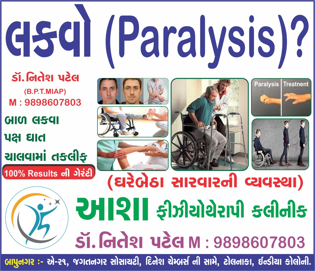
If you are searching Best Physiotherapy clinic for neuro in Ahmedabad, Here you will find best center for you. Physiotherapy treatment is an important part of Rehabilitation of neuro related condition such as Paralysis, Hemiplegia, Paraplegia.

Physiotherapy treatment are uses various exercise protocol and equipment to restore body movement and help you to recover from bed-ridden condition to normal.

In this article we will update list of Best Physiotherapy clinic who are experienced and qualified Physiotherapist.

Best Physiotherapy clinic for neuro in Bapunagar, Ahmedabad

Asha Physiotherapy Clinic

Physiotherapy Center Address:

A -21, Jagatnagar Part-1,Opp. Dinesh Chambers
India Colony Road,Nr. Tolnaka
Bapunagar,
Ahmedabad - 380024
Gujarat

Asha Physiotherapy clinic and neuro rehab center is the first Physiotherapy clinic in east Ahmedabad with all latest physiotherapy equipment with highly qualified and trained staff.

Home visit treatment facility is also available in east Ahmedabad for neuro related patient.

Dr. Bhavnaben Malavia - Physiotherapist

Dr. Nitesh Patel - Physiotherapist

(BPT, MIAP)

Dr. Niral Kathiriya - Physiotherapist
Dr. Nidhi Ribadiya - Physiotherapist
Dr.Neha Nai - Physiotherapist
Dr. Jyoti - Physical therapist
Dr. Dharti Patel - Physiotherapist

Clinic Timing:

  • Monday : Morning : 8:300 am to 1 :00 pm, Evening :4 :00 pm to 8:00pm.
  • Tuesday : Morning : 8:300 am to 1 :00 pm, Evening :4 :00 pm to 8:00pm.
  • Wednesday : Morning : 8:300 am to 1 :00 pm, Evening :4 :00 pm to 8:00pm.
  • Thursday : Morning : 8:300 am to 1 :00 pm, Evening :4 :00 pm to 8:00pm.
  • Friday : Morning : 8:300 am to 1 :00 pm, Evening :4 :00 pm to 8:00pm.
  • Saturday : Morning : 8:300 am to 1 :00 pm, Evening :4 :00 pm to 8:00pm.
  • Sunday : Morning : 8:300 am to 12 :00 pm, Evening : Holiday


Facility in Asha Physiotherapy center:

The center have all latest physiotherapy equipment and well-spaced and experienced staff for the Best Physiotherapy treatment.

Electrotherapy Equipment in The clinic:

Following are the list of Physiotherapy related equipment in Asha Physiotherapy clinic.

  • Short wave Diathermy (SWD)
  • Interferential Therapy (IFT)
  • Infra Red Therapy (IR)
  • Ultrasound Therapy (US)
  • Paraffin Wax Bath Therapy
  • Electrical Stimulation (ES)
  • Lumbar Traction cum Cervical Traction Machine
  • Ultraviolet Therapy (UVR)
  • Trans-cutaneous Electrical Nervous Stimulation (TENS)
  • Vibrator Machine
  • Treadmill

Mobile Physiotherapy Clinic Bapunagar

B-1, Jagatnagar Society, Opp.Shaktidhara Society
India Colony Road, Tollnaka, Bapunagar, Ahmedabad.

Dr Nitesh Patel – Physiotherapist.
Dr Vaishali Ladva – Physiotherapist
Dr Nidhi Ribadiya – Physiotherapist
Dr Priti Tiragar – Physiotherapist

Clinic time:

Morning Session: 8: 30 am to 1:00 pm
Evening Session: 4:00 pm to 8:00 pm

Samarpan Physiotherapy Clinic Bapunagar Branch

Chandragupt flat, Near Ramaji mandir
Near. Satsangi school, India colony road
Behind Nilkanth dairy
Bapunagar, Ahmedabad

Physiotherapist in the clinic:

Dr Nitesh Patel- Physiotherapist
Dr Vaishali Ladva- Physiotherapist
Dr Dharti Vaghasiya- Physiotherapist
Dr Aarti Solanki- Physiotherapist
Dr Niyati Prajapati- Physiotherapist

Time :

Morning Session: 8: 30 am to 1:00 pm
Evening Session: 4: 00 pm to 8:00 pm

website: https://samarpanphysioclinic.com

 Best Physiotherapy clinic for neuro in Nava Naroda Ahmedabad

 Samarpan Physiotherapy clinic Nava Naroda Nikol Road Branch

11, Vedant Bungalow, Opposite. Radhe -2 Bungalow,
Near. Fortune Circle, Nikol Nava Naroda Road,
New India colony road
Behind. Shalby Hospital
Nava Naroda, Ahmedabad.

Nearby location is Govardhan galaxy bungalow

Physiotherapists in the Clinic

Dr Varsha Patel – Physiotherapist
Dr Nitesh Patel – Physiotherapist
Dr Shivangi Chauhan– Physiotherapist
Dr Shivani Brahmabhatt – Physiotherapist
Dr. Pritiksha Patel – Physiotherapist

Time :

Morning Session: 8: 30 am to 1:00 pm
Evening Session: 4:00 pm to 8:00 pm

Best Physiotherapy clinic for neuro in Vastral, Ahmedabad

Samarpan Physiotherapy clinic Vastral Branch

B-04, Shivalik Bungalow, Near. Metro pillar no. 156
Near. Ratanpura gam, Madhav school road
Near. Vedant play school
Vastral, Ahmedabad

Nearby locations are Bhavani party plot and Nirant char rasta towards 156 pillar

Physiotherapist in the clinic:

  • Dr Nitesh Patel- Physiotherapist
  • Dr Falguni Luhar- Physiotherapist
  • Dr Srusti Vaghasiya- Physiotherapist
  • Dr Akshita Mehta- Physiotherapist
  • Dr. Payal Patel - Physiotherapist


Clinic Time :

Morning Session: 8: 30 am to 1:00 pm
Evening Session: 4:00 pm to 8:00 pm

Best Physiotherapy clinic for neuro in Amaraiwadi, Ahmedabad

Samarpan Physiotherapy clinic Amaraiwadi branch address


Bansidhar Society Behind Bansidhar Medical, Vastral Road,
Rabari Colony Cross Road,
near Metro Pillar No 77-78,
Opposite revabhai shopping center
Amraiwadi, Ahmedabad, Gujarat 380026

Nearby location are Sureliya char rasta towards Rabari colony char rasta

Dr. Nitesh Patel – Physiotherapist. Mo no: 09898607803
Dr. Komal Chauhan – Physiotherapist- Mo no: 06352845017
Dr. Mohit Vaghela – Physiotherapist
Dr. Rahul Rathod – Physiotherapist
Dr. Namrata Prajapati – Physiotherapist

Home Visit treatment facility for neuro rehabilitation:

Mostly neuro patient are bed-ridden and unable to go Physiotherapy clinic daily basis, so Home visit treatment is required until patient start walking, so physiotherapist from clinic visit patient home and according to condition given treatment. 

Home visit treatment fee varies on patient's condition and how far is the location. If you are also looking for Home visit treatment please call Dr. Nitesh Patel- Physiotherapist Contact no is 09898607803.

Thursday, 31 March 2022

Physiotherapy center near india colony, ahmedabad, gujarat


Paralysis treatment
Paralysis treatment

If you are in search for Best Physiotherapy center near India Colony Road Bapunagar area, then This article will inform you list of Best Physiotherapy center in your area.

Here we update list of Best Physiotherapy center located near India Colony Road Bapunagar, Our list is Base on Experience, Quality of treatment, Facility in the clinic and reviews available on Google.

List of Best Physiotherapy Center in India Colony Road Bapunagar:

Following are the List of Physiotherapy clinic where top listed will be in top position and are also updated accordingly.

  • Asha Physiotherapy center
  • Samarpan Physiotherapy center
  • Mobile Physiotherapy center
  • Jyoti Physiotherapy Weight Care & Fitness Center
  • Ashirvad Physiotherapy center
  • Dharti Physiotherapy center
  • Sanidhya Physiotherapy Fitness & Rehab center
  • Stuti Physiotherapy center
  • Shreeji Physiotherapy center
  • Aashray Pediatric NDT & SI center


Asha Physiotherapy Center:


Asha Physiotherapy clinic is started By Dr. Bhavnaben Malavia - Physiotherapist, First Physiotherapy clinic in Bapunagar and India Colony Road. Over the Time Clinic have developed all latest Physiotherapy Equipment and Facility and Now becoming one of the Highest Facility-wise and experienced clinic in Bapunagar area.

More than 31 years of Experienced Rehabilitation team gives you advanced Physiotherapy treatment option where you cannot find in other clinic.

Physiotherapy Center Address:

A -21, Jagatnagar Part-1,Opp. Dinesh Chambers
India Colony Road,Nr. Tolnaka
Bapunagar,
Ahmedabad - 380024
Gujarat.

Dr. Bhavnaben Malavia - Physiotherapist

Dr. Nitesh Patel - Physiotherapist

(BPT, MIAP)

Dr. Niral Kathiriya - Physiotherapist.

Dr.Neha Nai - Physiotherapist.
Dr. Jyoti - Physical therapist
Dr. Dharti Patel - Physiotherapist.

Clinic Timing :

  • Monday : Morning : 8:300 am to 1 :00 pm, Evening :4 :00 pm to 8:00pm.
  • Tuesday : Morning : 8:300 am to 1 :00 pm, Evening :4 :00 pm to 8:00pm.
  • Wednesday : Morning : 8:300 am to 1 :00 pm, Evening :4 :00 pm to 8:00pm.
  • Thursday : Morning : 8:300 am to 1 :00 pm, Evening :4 :00 pm to 8:00pm.
  • Friday : Morning : 8:300 am to 1 :00 pm, Evening :4 :00 pm to 8:00pm.
  • Saturday : Morning : 8:300 am to 1 :00 pm, Evening :4 :00 pm to 8:00pm.
  • Sunday : Morning : 8:300 am to 12 :00 pm, Evening : Holiday


Facility in Asha Physiotherapy center:

The center have all latest physiotherapy equipment and well-spaced and experienced staff for the Best Physiotherapy treatment.

Electrotherapy Equipment in The clinic:

Following are the list of Physiotherapy related equipment in Asha Physiotherapy clinic.

  • Short wave Diathermy (SWD)
  • Interferential Therapy (IFT)
  • Infra Red Therapy (IR)
  • Ultrasound Therapy (US)
  • Paraffin Wax Bath Therapy
  • Electrical Stimulation (ES)
  • Lumbar Traction cum Cervical Traction Machine
  • Ultraviolet Therapy (UVR)
  • Trans-cutaneous Electrical Nervous Stimulation (TENS)
  • Vibrator Machine
  • Treadmill

Samarpan physiotherapy clinic Bapunagar Branch:

Samarpan Physiotherapy clinic is started in 2005, By Dr. Nitesh Patel - Physiotherapist is also well-updated Best clinic in East Part of Ahmedabad, Samarpan Physiotherapy clinic have 4 branches  located mainly in East Part of Ahmedabad.

Dr. Nitesh Patel is a one of the Best Physiotherapist running more than Five Branches in East Ahmedabad, Clinic is located in Amaraiwadi, Vastral, Nava Naroda and India Colony Road Bapunagar area.

Bapunagar Physiotherapy center Address:

Samarpan Physiotherapy Clinic
Chandragupt Flat, Near.Ramji Mandir
Behind Nilkanth Dairy
Near. Satsangi School
India Colony Road
Bapunagar, Ahmedabad.

Dr. Nitesh Patel – Physiotherapist.
Mo :  9898607803

Dr. Vaishali Ladva – Physiotherapist.
Dr.Aarti Solanki – Physiotherapist
Dr. Dharti Vaghasiya - Physiotherapist.

Timing :

Morning : 8 : 30 am to 1 : 00 pm
Evening : 4 : 00 pm to 8 : 30 pm
Sunday : 9 : 00 am to 12: 00 pm

Vastral Physiotherapy Center Branch Address: Nirant Char Rasta

Samarpan Physiotherapy Fitness and Rehabilitation clinic.
B-04, Shivalik Bunglow, Near. Metro Pillar No. 156
Madhav School Road
Near. Ratanpura Gam ,
Vastral.

DR. NITESH PATEL: ( Physiotherapist )

M  : 9898607803
    : 7777976968

Dr. Ruchita Thakkar – Physiotherapist.
Dr. Akshita Mehta - Physiotherapist.
Dr. Poorva Patel - Physiotherapist
Dr. Falguni Patel - Physiotherapist.

Timing :

Morning :8 :30 am to 1 :00 pm
Evening : 4 :00 pm to 8 :00 pm

Sunday : 9 :00 am to 12:00 pm

Nava Naroda Physiotherapy Clinic Address:

Samarpan Physiotherapy Clinic

11, Vedant Bunglow, Opp.Radhe-2 Bunglow,
Near. Haridarshan Char Rasta, Behind.Shalby Hospital
Near. Fortune Circle, New India Colony Road
Sardar Patel Road, Nava Naroda, Ahmedabad.

Dr. Nitesh Patel – Physiotherapist.
Mo No. : 09898607803

Dr. Varsha Patel – Physiotherapist.
Dr. Dharti Patel - Physiotherapist.
Dr. Pankati Patel – Physiotherapist.

Timing :

Morning : 8 :30 am to 1 : 00 pm
Evening : 4 :00 pm to 8 : 00 pm
Sunday : 9 :00 am to 12:00 pm

Amaraiwadi Physiotherapy Clinic Address : Near Rabari Colony Char Rasta

Samarpan Physiotherapy Clinic

Bansidhar Society,Behind.Bansidhar Medical store
Vastral Road, Rabari Colony Char Rasta
Amaraiwadi,Ahmedabad

Dr. Komal Chauhan – Physiotherapist.

Mo :  6352845017
      : 7285084764

Dr. Mohit Vaghela – Physiotherapist.
Dr. Namrata Prajapati – Physiotherapist
Dr. Khusbu Patel – Physiotherapist.

Timing :

Morning : 8 :00 am to 1 : 00 pm
Evening : 4 :00 pm to 8 : 30 pm

Sunday : 9 :00 am to 12: 00 pm

Mobile Physiotherapy Clinic:

B-, Jagatnagar Society, Opp.Shaktidhara Society
India Colony Road, Tollnaka, Bapunagar, Ahmedabad.

Dr. Nitesh Patel – Physiotherapist.
Dr. Vaishali Ladva – Physiotherapist
Dr. Pal Rawal – Physiotherapist
Dr.  – Physiotherapist

Jyoti Physiotherapy clinic:

This clinic is started by Dr. Sandip Chawda - Physiotherapist.

Clinic Address:

B-69,70,71, First Floor, Sardar Patel Mall, Nikol Gam Rd, Khodiar Nagar,
Ahmedabad, Gujarat 382350

Clinic Timings :
Monday to Saturday : 9am to 8pm

Mo No : 09879310606

Ashirvad Physiotherapy Clinic

This clinic is started by Nitinbhai is now shifted to Canada and Now clinic is running by Dr.Suresh Meghwal - Physiotherapist.

Ashirvad Physiotherapy Centre, Shastri Nagar, Bapunagar, Ahmedabad, Gujarat - 380038

Dharti Physiotherapy Center

Clinic Address :

Shop 8, Shilp Residency Ground Floor, Cross Rd, Hirawadi, Ahmedabad, Gujarat 380024
Ahmedabad.

Clinic timing :

Morning : 9:30am–1pm
Evening : 4:30–9pm

Sanidhya Physiotherapy Fitness & Rehab Center

 A/44, Shaktidhara Society, Opposite Shayano Arcade, Near Dinesh Chamber, Tollnaka, Bapu Nagar, Bapu Nagar, Ahmedabad, Gujarat 380024
Contact no : 079 6570 0264

Stuti Physiotherapy Center:

Shyam Shikhar Complex, Opp. Poojan Apartment,Behind, Tolnaka, Bapunagar, Ahmedabad, Gujarat 380024.

Shreeji Physiotherapy center

7, Shaktinagar Society, Anupam Colony, Bapunagar, Ahmedabad, Gujarat 382350

Dr. Bhavin Kaswala - Physiotherapist.

Aashray Pediatric NDT & SI Center

Devraj Mall, 307, India Colony Rd, Bapunagar, Ahmedabad, Gujarat 382350

Thursday, 17 February 2022

Paralysis treatment in Bapunagar, Ahmedabad

Paralysis treatment in Bapunagar, Ahmedabad
Paralysis treatment in Bapunagar

 If you are looking for advanced treatment of Paralysis in Bapunagar area, Asha Physiotherapy clinic and Neuro Rehabilitation center is the best option for you.

Asha Physiotherapy clinic offers you best treatment option for Paralysis where experienced Physiotherapist and staff give best quality treatment with result oriented treatment plan. This clinic is started in 1989 by Dr. Bhavnaben Malvia with purpose of giving best to society in the field of Physiotherapy.

Asha Physio clinic have all advanced equipment related to Physiotherapy that are also useful for Paralysis.

Paralysis related condition such as Hemiplegia, Paraplegia, Quadruplegia, Cerebral Palsy, stroke, GBS etc. where personal treatment is required. In Asha Clinic experienced staff working since more than 30 years giving best of their possible.

Home Visit Treatment facility:

If you are unable to visit clinic mainly due to bed-ridden condition, Physiotherapist from Asha clinic come to you at your home and giving treatment accordingly. Home visit treatment facility is available in all area of East Ahmedabad such as Bapunagar, India colony road, Vastral, Amaraiadi, Odhav, Nava Naroda, Nikol, Rakhial, Saraspur, Maninagar, CTM etc.

Home Visit Treatment

Asha Physiotherapy Clinic:

Physiotherapy Center Address:

A -21, Jagatnagar Part-1,Opp. Dinesh Chambers
India Colony Road,Nr. Tolnaka
Bapunagar,
Ahmedabad - 380024
Gujarat.

Dr. Bhavnaben Malavia - Physiotherapist

Dr. Nitesh Patel - Physiotherapist

(BPT, MIAP)

Dr. Niral Kathiriya - Physiotherapist
Dr. Nidhi Ribadiya - Physiotherapist
Dr.Neha Nai - Physiotherapist
Dr. Jyoti - Physical therapist
Dr. Dharti Patel - Physiotherapist

Clinic Timing:

  • Monday : Morning : 8:300 am to 1 :00 pm, Evening :4 :00 pm to 8:00pm.
  • Tuesday : Morning : 8:300 am to 1 :00 pm, Evening :4 :00 pm to 8:00pm.
  • Wednesday : Morning : 8:300 am to 1 :00 pm, Evening :4 :00 pm to 8:00pm.
  • Thursday : Morning : 8:300 am to 1 :00 pm, Evening :4 :00 pm to 8:00pm.
  • Friday : Morning : 8:300 am to 1 :00 pm, Evening :4 :00 pm to 8:00pm.
  • Saturday : Morning : 8:300 am to 1 :00 pm, Evening :4 :00 pm to 8:00pm.
  • Sunday : Morning : 8:300 am to 12 :00 pm, Evening : Holiday


Facility in Asha Physiotherapy center:

The center have all latest physiotherapy equipment and well-spaced and experienced staff for the Best Physiotherapy treatment.

Electrotherapy Equipment in The clinic:

Following are the list of Physiotherapy related equipment in Asha Physiotherapy clinic.

Short wave Diathermy (SWD)
Interferential Therapy (IFT)
Infra Red Therapy (IR)
Ultrasound Therapy (US)
Paraffin Wax Bath Therapy
Electrical Stimulation (ES)
Lumbar Traction cum Cervical Traction Machine
Ultraviolet Therapy (UVR)
Trans-cutaneous Electrical Nervous Stimulation (TENS)
Vibrator Machine
Treadmill

વજન ઘટાડવાના સરળ ઉપાયો: એક સંપૂર્ણ ગુજરાતી માર્ગદર્શિકા

વજન ઘટાડવું એ માત્ર શારીરિક દેખાવ સુંદર બનાવવાની બાબત નથી, પરંતુ તે એક સ્વસ્થ, રોગમુક્ત અને ઊર્જાસભર જીવન જીવવાની દિશામાં સૌથી મહત્વનું કદમ ...